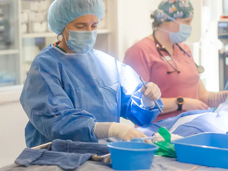
Cranial Cruciate Disease

The cranial cruciate ligament (CrCL) is one of the four ligaments within the knee and is the most important stabilizer of the stifle joints in dogs. Injury to the CrCl is the most common orthopedic disease that we see in the veterinary field. This is analogous to anterior cruciate ligament (ACL) in humans. This ligament connects the femur to the tibia and prevents instability as the dog uses its leg.


Unlike human athletes, rupture of the CrCL in dogs is rarely from a traumatic injury. This is actually more of a degenerative condition meaning that the ligament is degenerating (weakening) over time. This can be secondary to genetic, conformation, or immune-mediated causes within the joint. Once the ligament is weakened, it may partially or completely rupture following any activities such as jumping or running.  CrCL rupture is the most common cause of stifle (knee) problems in all dogs. About 50% of dogs will rupture both CrCl’s within 1-2 years of each other. Dogs will frequently also tear the meniscus at the time of the cruciate rupture.


Dogs present with a significant lameness of the affected leg. Once the cruciate ligament tears, the joint will become swollen which adds to the pain and lameness. There will be thickening of the joint and then arthritis develops. As lameness continues, muscle atrophy occurs. If the lameness becomes chronic, the joint can start to develop scar tissue and lose range of motion and normal function. Dogs may have trouble getting up or lying down and can have worse lameness after exercise. Some patients will have tears on both hind legs and may be unable to walk at all without significant assistance.


Diagnosis of cranial cruciate disease includes physical examination with an orthopedic specialist. A physical exam will reveal swelling in the stifle, pain on range of motion, thickening of the joint, and lameness +/- muscle atrophy. Tests including cranial drawer and tibial thrust will be performed to evaluate for any instability of the stifle. Radiographs do not show the actual CrCl tear, but they support the diagnosis by showing joint effusion as well as secondary changes such as arthritis or degenerative joint disease.


The definitive diagnosis of cranial cruciate disease will be performed via arthroscopy or arthrotomy during surgical stabilization. Surgical stabilization is the treatment of choice for cranial cruciate ruptures in dogs. There have been many different surgical procedures described in veterinary medicine for the stabilization of cranial cruciate ruptures. The most successful include tibial plateau leveling osteotomy (TPLO), lateral suture (LFS) and tibial tuberosity advancement (TTA). Your orthopedic surgeon will discuss surgical recommendations and options with you.


By far the most commonly performed technique is the TPLO. This is considered by many surgeons to be the most successful surgical method for stabilization of the stifle for cranial cruciate disease in all dogs. During TPLO, the biomechanics of the stifle joint are altered to compensate for the cruciate rupture. (there is not a good technique in veterinary medicine for replacement of the CrCl with a graft or suture). The tibia in dogs has a downward slope and the intact CrCL prevents the femur from sliding down the slope of the tibia when dogs are bearing weight. In a stifle with a ruptured CrCL, the femur slides down the slope. The TPLO prevents this forward rotation from occurring by flattening the tibial plateau. A plate is placed to secure the osteotomy. TPLO surgery carries an excellent prognosis for returning your dog to its normal activities including walking running, playing, and jumping without pain and lameness.