Facts about Glaucoma
By Dr. Rachel Mathes, DVM, MS, Diplomate ACVO
- Secondary glaucoma more common than primary glaucoma
- Females overrepresented 2:1 for primary glaucoma
- Primary glaucoma typically associated with iridocorneal angle closure and increased intraocular pressure during middle age (6-8 years old)
- Canine glaucoma tends to be aggressive
- Feline glaucoma is almost exclusively secondary
- Concurrent institution of prophylactic therapy for the contralateral eye should be instituted if an eye is enucleated for primary glaucoma
- Imperative to address IOP spikes immediately as they may quickly cause complete blindness
Glaucoma is a term used to describe a group of diseases that cause elevated, often severely, intraocular pressure. This condition is very painful and treatment is aimed not only at preservation of vision, but also for pain management. The pain of glaucoma may be referred pain (migraine headache) and patients may exhibit discomfort by decreased activity, increased sleeping or subtle changes in behavior.1 The signs of pain may not even be noted by the owner until the discomfort is treated, at which time the patient may be noted to return to normal activity or “act like themselves again.” Primary, or breed-related, glaucoma in dogs is most commonly due to closure of the aqueous drainage angle or Primary Angle Closure Glaucoma (PACG).1 Females are approximately twice as likely to develop PACG compared to males.2 PACG has different features for different canine breeds, however, the end result is failure of normal aqueous outflow, causing significant intraocular pressure elevation. Acute primary glaucoma may often be treated medically or surgically if addressed immediately, even if there is vision loss at the time of increased intraocular pressure.1,3,4 Chronic primary glaucoma causes extensive intraocular damage and blindness. Therapy is aimed at preventing intraocular pressure spikes, decreasing intraocular pressure, and maintaining functional vision.4-7 This therapeutic goal is rarely achieved long term with medication alone and surgical intervention is almost always necessary.5 To preserve functional vision, glaucoma surgery is usually recommended early in the disease course due to the aggressive nature of this disease. Typical surgeries performed include laser cyclophotocoagulation (ciliary body destruction) and anterior chamber valve placement.5 The combination of these surgeries resulted in good control of the intraocular pressure in 76% of cases.5 In cases of irreversibly blind globes, more permanent salvage procedures are recommended as glaucoma is painful (enucleation or an intrascleral prosthesis).
Secondary glaucoma results from other underlying ocular pathology. The most common causes are lens luxations (most often seen in Terrier breeds), uveitis, and cataracts. Treatment is aimed at reducing the intraocular pressure and addressing the underlying cause of pressure elevation. Topical glaucoma therapy should be limited, in cases of secondary glaucoma, to medications that do not exacerbate pre-existing ocular disease. Topical carbonic anhydrase inhibitors (e.g. dorzolamide, brinzolamide) or topical beta blockers (e.g. timolol) are recommended for primary or secondary glaucoma. Prostaglandin analogs (e.g. latanoprost), while often the first line of therapy for primary glaucoma, should be avoided for secondary glaucoma therapy as they may exacerbate pre-existing ocular disease.
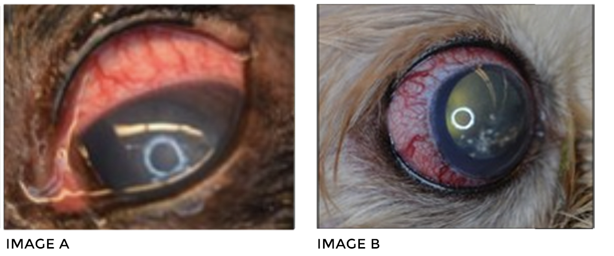
 A patient with acute glaucoma is depicted (A). Note the severe scleral injection and corneal edema. Acute glaucoma must be addressed immediately in order to preserve vision and treat patient discomfort. A patient with chronic glaucoma is depicted (B) Note the significant buphthalmos (globe enlargement), scleral injection, and corneal edema. This eye is irreversibly blind, but is quite painful. Therapy is aimed at providing long-term patient comfort. Prophylactic therapy should always be instituted in the contralateral eye if an eye is removed for intractable, primary glaucoma.
REFERENCES
- Reinstein S, et al. Canine glaucoma: pathophysiology and diagnosis. Compend Contin Educ Vet. 2009:10;450-2.
- Tsai S, et al. Gender differences in iridocorneal angle morphology: a potential explanation for the female predisposition to primary angle closure glaucoma in dogs. Vet Ophthalmol. 2012:15S1;60-3.
- Scott E, et al. Early histopathologic changes in the retina and optic nerve in canine primary angle-closure glaucoma. Vet Ophthalmol. 2013:16S1;79-86.
- Dees D, et al. Efficacy of prophylactic antiglaucoma and anti-inflammatory medications in canine primary angle-closure glaucoma: a multicenter retrospective study (2004-2012). Vet Ophthalmol. 2013:5.
- Sapienza J, et al. Combined transscleral diode laser cyclophotocoagulation and Ahmed gonioimplantation in dogs with primary glaucoma: 51 cases (1996-2004). Vet Ophthalmol. 2005:8:121-7.
- Miller P, et al. The efficacy of topical prophylactic antiglaucoma therapy in primary closed angle glaucoma in dogs: a multicenter clinical trial. J Am Anim Hosp Assoc. 2000:36:431-8.
- Willis A, et al. Advances in topical glaucoma therapy. Vet Ophthalmol. 2002:5;9-17.