About MRI and CT in Veterinary Medicine
Why use MRI in neurology?
- In both human and veterinary neurology, magnetic resonance imaging (MRI) is the gold standard imaging modality.
- MRI uses a strong magnetic field to manipulate the position of protons. Protons help make up atoms, which are the building block for cells.
- The MR scanner applies a series of radiofrequency pulses. This causes the protons to move in a unique way, depending on the type of cells they compose (water, fat, etc). This information is delivered to the computer which can build detailed 2D pictures of the targeted area.
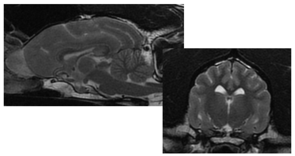
- This unique ability to differentiate fat from water makes MRI superior in assessing soft tissue detail.
- Neurologists use MRI to view the patient’s nervous system (brain, spinal cord, and peripheral nerves). This amazing soft tissue detail gives a better understanding of a patient’s nervous system, why the patient may be having neurological signs, and allows the neurologist to make the most appropriate treatment and testing recommendations.
What is the difference between an MRI and CT?
- A CT scan (which stands for computed tomography) is another advanced imaging modality used in both human and veterinary specialty medicine.
- A CT uses high-energy electromagnetic radiation (X-rays) to create cross sectional images of the body. This modality creates 2D and 3D images similar to radiographs.
- CT imaging is valuable when assessing bone, monitoring for bleeding in the brain or abdomen, identifying blood vessels, and scanning for certain types of neoplasia (cancer).
- A neurologist may recommend a combination of MRI and CT, depending on the history and examination of a neurological patient.