Facts About Limbal Melanomas
By Rachel Mathes, DVM, MS, Diplomate ACVO
- Benign, slow-growing tumors in dogs and cats
- Very responsive to a variety of therapies
- Low rate of metastasis
- Low rate of recurrence with treatment
- Early referral recommended
Limbal melanomas are benign, slowly growing tumors of limbal melanocytic origin. These tumors typically occur along the dorsomedial to ventrolateral limbal arc of the globe at the corneal and scleral junction.1 A bimodal age distribution has been described with a peak occurrence at 3-4yrs of age and 7-10yrs of age in dogs (1,2). These tumors are thought to have an inherited basis as Golden Retrievers are four times more likely and Labradors are three times more likely to develop this compared to other breeds (1). Though benign, limbal melanomas may become globe-threatening with growth due to local invasion. Often, secondary keratitis with corneal lipid deposition will occur as a result of the tumor presence (2,3). While these tumors are less common in cats, they may occur and have a similar clinical course as canine melanomas (3). Feline and canine limbal melanomas are amenable to a variety of therapies including surgical debulking with a combined keratectomy and sclerectomy, cryotherapy, laser photocoagulation, radiation, and surgery with homologous and autologous grafting (2,3,4). In one recent study, only 1 out of 30 tumors recurred after a combination therapy protocol (2). In addition, the incidence of side effects from surgery was low with less than 10% of complications being potentially globe threatening (2). After laser photocoagulation, a low recurrence was reported with 1 out of 15 masses recurring at 3 months and 2 out of 15 recurring at one year. Twelve of the fifteen tumors did not recur (3).
Long-term control and vision are attainable and reasonable goals with appropriate therapy. Early referral is recommended to preserve vision and globe integrity.
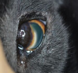
A dorsomedial limbal melanoma in the left eye is pictured. The mass is heavily pigmented, raised, irregular, and well-demarcated with an arc of white corneal lipid at the leading edge.